The Complete Guide to Mole (Nevus) Removal
— Principles of Selective Tissue Ablation with CO2 and Erbium Lasers and Scarless Treatment Strategies
Moles (nevi) on the face or body are a common cause of cosmetic concerns. Moles are benign skin lesions characterized by localized proliferation of melanocytes. Depending on their size, depth, and type, they can sometimes represent more than just a cosmetic issue, requiring diagnosis and monitoring. However, mole removal is often perceived as a 'simple procedure,' leading to a significant number of cases experiencing side effects such as recurrence or scarring.
At BA Clinic, we accurately assess the histological type and depth of moles before designing a personalized treatment strategy using CO2 and Erbium (Er:YAG) lasers to minimize scarring and reduce recurrence rates.
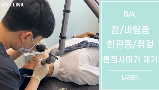
1. Medical Classification of Moles (Nevi) — Why Understanding 'Types' is Crucial
Moles are not a single entity. Histologically, they are broadly classified into three types based on the 'location (layer)' of melanocytes, and this classification dictates the laser treatment strategy.
- Junctional Nevus
Melanocytes are located at the dermoepidermal junction. These are typically flat, uniformly brown, and superficial, responding well to laser treatment. They can often be removed in 1-2 sessions.
- Compound Nevus
Melanocytes are distributed at the dermoepidermal junction and in the upper dermis. These may be slightly raised, and residual cells within the dermis can lead to recurrence even after superficial pigment removal. This type requires a phased treatment approach.
- Intradermal Nevus
Melanocytes are located deep within the dermis. These appear as skin-colored or light brown hemispherical bumps. Due to their depth, precise control of tissue ablation is necessary during laser treatment to prevent scarring.
Other types, such as Blue Nevus, Giant Congenital Nevus, and Dysplastic Nevus, require observation or biopsy first. Clinical evaluation by a specialist is essential before laser removal.
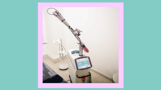
2. Principles of Mole Removal Lasers — CO2 Laser vs. Erbium (Er:YAG) Laser
The two most commonly used lasers for mole removal are the CO2 (Carbon Dioxide) laser and the Erbium (Er:YAG) laser. Both lasers work by vaporizing tissue using water as the chromophore. However, there are critical differences in their wavelengths and thermal diffusion characteristics.
- CO2 Laser (Wavelength 10,600nm)
It has a high water absorption coefficient, providing strong tissue vaporization capabilities. Simultaneously, thermal diffusion occurs in surrounding tissues. This thermal effect is advantageous for hemostasis and facilitates clear visualization during the removal of deep lesions. However, excessive thermal damage can lead to hypertrophic scars or post-inflammatory hyperpigmentation (PIH), making precise control of energy density and exposure time essential.
- Erbium (Er:YAG) Laser (Wavelength 2,940nm)
Its water absorption rate is about 12-18 times higher than the CO2 laser, allowing for precise removal of very thin layers of tissue (approximately 5-20μm). Minimal thermal diffusion results in less damage to surrounding normal tissue and faster recovery. However, its hemostatic effect is weaker, potentially leading to bleeding, and multiple passes may be required for deep lesions.
At BA Clinic, we selectively apply or combine these two lasers depending on the depth, size, and location of the mole to achieve maximum lesion removal with minimal tissue damage.

3. Causes of Scarring After Mole Removal — The Science of Depth Control
The most common cause of scarring after mole removal is 'excessive depth of tissue ablation'. The skin consists of the epidermis (approx. 0.1mm) and dermis (approx. 1-4mm). If ablation is limited to the papillary dermis (upper dermis), re-epithelialization from skin appendages (hair follicles, sweat glands) occurs smoothly, leading to scar-free healing.
However, if tissue is destroyed beyond the reticular dermis (mid-dermis), the rate of re-epithelialization slows down, and excessive collagen production by fibroblasts can lead to hypertrophic or atrophic scars.
This is precisely why mole removal is not a 'simple procedure.' The practitioner must make real-time judgments on the following:
- The current depth reached by the laser (epidermis vs. papillary dermis vs. reticular dermis)
- Whether residual melanocytes can be visually assessed
- The risk of scarring if additional passes are performed
- Whether to aim for complete removal in one session or opt for staged treatment
At BA Clinic, prioritizing 'scarless results over complete removal,' we adopt a staged approach for lesions of questionable depth. We ablate only to a safe depth in the first session and re-evaluate after 4-8 weeks to determine if further treatment is needed. This conservative strategy yields the best long-term results.

4. Recurrence Mechanism — Why Do Moles Come Back?
Recurrence after mole removal is one of the most common patient complaints. Medically, recurrence is explained by two main mechanisms:
- Recurrence from Residual Melanocytes
This occurs when surface pigment is removed by the laser, but residual melanocytes deep in the dermis reactivate and produce pigment again due to external stimuli like UV radiation. This is particularly common in compound or intradermal nevi and is a natural phenomenon when the lesion's depth exceeds the safe ablation range, rather than a failure of the procedure.
- Post-Inflammatory Hyperpigmentation (PIH)
Often, temporary post-inflammatory hyperpigmentation is mistaken for recurrence. This is common in Asian skin (Fitzpatrick Type III-IV) and typically resolves naturally within 3-6 months with UV protection and proper care.
At BA Clinic, we evaluate lesion depth and borders using dermoscopy before treatment and plan staged treatment for types with a high recurrence potential. We also provide a systematic post-treatment care protocol, including UV protection and topical medication, to prevent post-inflammatory hyperpigmentation.
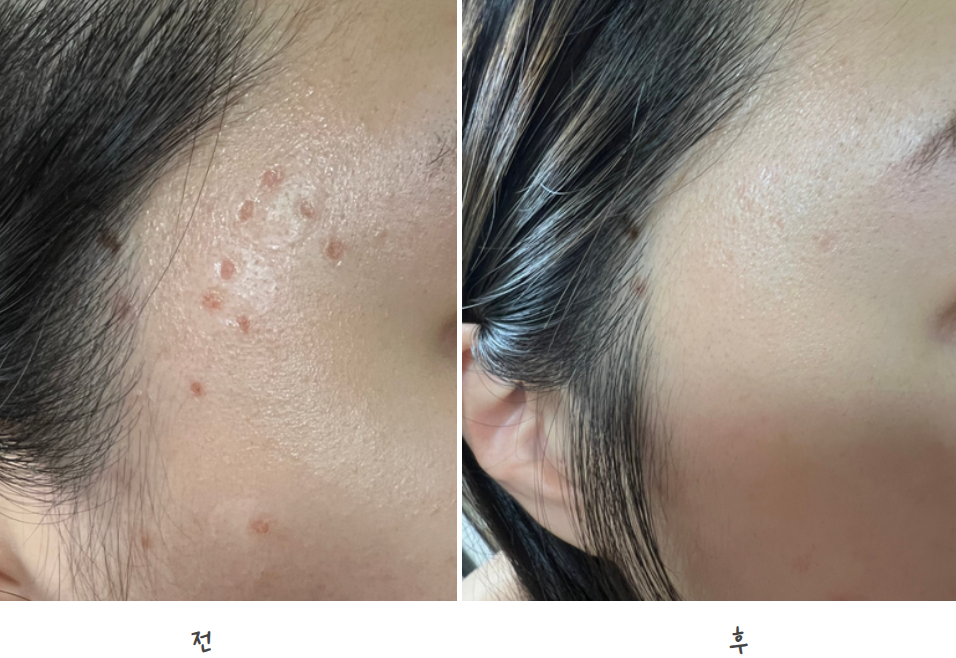
5. Precautions by Area — Mole Removal on the Face and Body Differs
Even moles of the same size require different treatment approaches depending on their location.
- Around the Nose and Mouth
Sebaceous glands are active, and the skin is relatively thicker, leading to faster healing. However, scars are more noticeable around the nose, so energy density is set conservatively.
- Eyelids and Around the Eyes
The skin is very thin (approx. 0.5mm) and densely vascularized, requiring precise energy control. The precise ablation capability of the Erbium laser is advantageous for this area.
- Forehead and Temples
These areas have a relatively broad and flat skin structure, allowing for fast and effective removal with a CO2 laser. However, as they are easily exposed to sunlight, post-treatment hyperpigmentation management is crucial.
- Torso and Limbs
Compared to the face, the density of skin appendages is lower, resulting in slower re-epithelialization and a relatively higher risk of scarring. Areas like the chest, shoulders, and back are prone to keloid scarring, so assessing the patient's tendency for keloid formation is essential before treatment.
6. Post-Treatment Care Dictates the Outcome
Proper post-laser mole removal care is as crucial as the procedure itself for preventing scars and recurrence.
- Maintain a Moist Environment: Applying a hydrocolloid dressing speeds up re-epithelialization by up to 40% and inhibits scar formation. It is recommended to maintain this for 7-14 days post-procedure.
- Sun Protection: Exposure of the treated area to UV radiation significantly increases the risk of PIH. It is essential to apply SPF 50+ sunscreen frequently for at least 3 months and avoid direct sunlight.
- Scab Management: Prematurely removing scabs can disrupt re-epithelialization, leading to atrophic scars. It is best to let them fall off naturally.
- Recurrence Monitoring: Evaluate for recurrence 4-8 weeks post-treatment and plan further treatment if necessary. Early detection allows for clean retreatment with minimal energy.
7. Mole Removal at BA Clinic
Mole removal is not merely 'firing a laser'; it is an integrated medical procedure that involves understanding the lesion's histology, selecting appropriate lasers and energy settings, controlling depth in real-time, and providing systematic post-treatment care.
At BA Clinic, we perform mole removal based on the following principles:
- Pre-treatment evaluation of lesion depth and type using dermoscopy.
- Customized selection and combination of CO2 and Erbium lasers based on the lesion.
- Conservative depth control prioritizing 'scarless results.'
- Staged treatment plans for types with high recurrence potential.
- Post-treatment protocols to prevent hyperpigmentation and minimize scarring.
The difference in results, even for removing a single mole, stems from the practitioner's anatomical understanding and experience. If you are seeking treatment for a mole you've long been concerned about, starting with an accurate diagnosis, we invite you to consult with us at BA Clinic.
BA Clinic | 4F, 226, Daedeok-daero, Seo-gu, Daejeon Metropolitan City
Appointment Booking: 0507-1342-7053 | KakaoTalk Consultation Available
Weekdays 10:00~20:00 / Weekends 10:00~16:00 (Open through lunch)